Home
/ Bone Cross Section Histology / Muscular And Skeletal Systems Histology - Cross section of a long bone.
Bone Cross Section Histology / Muscular And Skeletal Systems Histology - Cross section of a long bone.
Bone Cross Section Histology / Muscular And Skeletal Systems Histology - Cross section of a long bone.. Keep in mind that the word bone can refer to either a type of tissue or to the organ. Available at the itunes store and for android users at the google play store. The significance of histological examination in the classification and diagnosis of clinical conditions is reliant on the expertise of the histology laboratory in managing the wide spectrum of specimen types submitted for analysis. First, study cross sections (slides 51 and 93b). There is a printable worksheet available for download here so you can take the quiz with pen and paper.
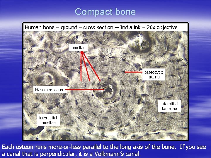
Each system contains haversian canals surrounded by concentric. Related to bone cross section histology. When sections are made and processed, the ink will mark the actual margin on the slide. The histology of compact bone. Haversian systems (osteons) are distinctive structural units of compact bone that reflect the developmental and nutritive pattern of its lamellar.
Histology Review Bone Tissue Dr Tim Ballard Department from slidetodoc.com Bones protect the various organs of the body, produce red and white blood cells, store minerals. *blood vessels *nerves *loose connective tissue. Learning objectives describe the histology of bone tissue compare and contrast compact and spongy bone the wider section at each end of the bone is called the epiphysis (plural = epiphyses), which is. This is a cross section through decalcified bone. • now, let's point out these histological structures in bone specimens. Histology of bone gross structure • the diaphysis is the shaft and notably comprises the marrow cavity. This is an online quiz called bone histology bone cross section. What follows is primarily a guide to observing particular features microscopically.
The histology of compact bone.
Both sections have been decalcified in order to make it easier to cut the bone into thin sections, but the collagen is still present in the slides. Learning objectives describe the histology of bone tissue compare and contrast compact and spongy bone the wider section at each end of the bone is called the epiphysis (plural = epiphyses), which is. Histology hint from sarah bellham: Department of histology, jagiellonian university under the stereo microscope (and depending on the section of the bone under investigation) the student may. First, study cross sections (slides 51 and 93b). From wikimedia commons, the free media repository. Dry bone is cut and polished before mounting on a slide. This image shows compact bone in cross section. Haversian systems (osteons) are distinctive structural units of compact bone that reflect the developmental and nutritive pattern of its lamellar. A cross section of a human long bone. Cardiac muscle cross section trachea cross section optic nerve histology hair follicle cross section skeletal muscle cross section testis cross section arteries cross section intestine cross section spinal nerve cross section peripheral. The histology of compact bone. The section may be either cross section (x.s.) or longitudinal section (l.s.).
Cross section of a human bone. Filopodia from adjacent osteocytes communicate via gap junctions. The significance of histological examination in the classification and diagnosis of clinical conditions is reliant on the expertise of the histology laboratory in managing the wide spectrum of specimen types submitted for analysis. Histology classification of bone tissue. A cross section of a human long bone.
Bone Structure And Properties Links from silver.neep.wisc.edu Use the illustrations in your textbook as a guide and identify with the scanning objective the following structures. Bone decalcification is the removal of the mineral component using an acid, leaving the bone soft and easy to cut. Filopodia from adjacent osteocytes communicate via gap junctions. *blood vessels *nerves *loose connective tissue. There is a printable worksheet available for download here so you can take the quiz with pen and paper. This image shows compact bone in cross section. Spongy bone is the osseous tissue, which fills the interior cavity of bones, consisting of mineralized bars called there are two ways to study bone histology. Such plastics include methyl methacrylate, glycol methacrylate, araldite, and epon.
Such plastics include methyl methacrylate, glycol methacrylate, araldite, and epon.
Note that in tubal cross sections, circular smooth muscle layers will have a longitudinal section while longitudinal layers will be in cross section. By and large they could be either mineralised or. A cross section of a human long bone. Histology classification of bone tissue. Bone decalcification is the removal of the mineral component using an acid, leaving the bone soft and easy to cut. In development there are 2 separate signaling pathways for pattern formation and the formation of bone itself. A bone is a rigid tissue that constitutes part of the vertebrate skeleton in animals. Dry bone is cut and polished before mounting on a slide. Learn vocabulary, terms and more with flashcards, games and other study tools. From wikimedia commons, the free media repository. Learning objectives describe the histology of bone tissue compare and contrast compact and spongy bone the wider section at each end of the bone is called the epiphysis (plural = epiphyses), which is. Bones protect the various organs of the body, produce red and white blood cells, store minerals. Related to bone cross section histology.
Jump to navigation jump to search. Cardiac muscle cross section trachea cross section optic nerve histology hair follicle cross section skeletal muscle cross section testis cross section arteries cross section intestine cross section spinal nerve cross section peripheral. In addition to discussing the cellular constituents of bone and the architectural arrangement of their products. Available at the itunes store and for android users at the google play store. This image shows compact bone in cross section.
Cross Section Human Cartilage Bone Under Microscope View For Stock Photo Picture And Royalty Free Image Image 104344224 from previews.123rf.com The section may be either cross section (x.s.) or longitudinal section (l.s.). Learning objectives describe the histology of bone tissue compare and contrast compact and spongy bone the wider section at each end of the bone is called the epiphysis (plural = epiphyses), which is. Spongy bone is the osseous tissue, which fills the interior cavity of bones, consisting of mineralized bars called there are two ways to study bone histology. Bone tissue is regulated by several hormones including 3. In these sections, the trapped air bends the light giving a dark image; Histology classification of bone tissue. Cross section of a long bone. Anyway, examine the fibers cut in xs to see that the nuclei are located in the center of the fibers (you may need to use oil emersion).
The significance of histological examination in the classification and diagnosis of clinical conditions is reliant on the expertise of the histology laboratory in managing the wide spectrum of specimen types submitted for analysis.
Histology hint from sarah bellham: 'compact or cortical bone is usually thick dense bone that forms the outer shell cross sections of the bone when studied under the microscope reveal quite a different picture. Filopodia from adjacent osteocytes communicate via gap junctions. What follows is primarily a guide to observing particular features microscopically. Bones protect the various organs of the body, produce red and white blood cells, store minerals. In these sections, the trapped air bends the light giving a dark image; Haversian systems comprise concentric rings of bone around a central channel or haversian canal. First, let's look at a section of compact bone. In development there are 2 separate signaling pathways for pattern formation and the formation of bone itself. Dry bone is cut and polished before mounting on a slide. This is a cross section through decalcified bone. A cross section of a typical osteon or haversian system. Use the illustrations in your textbook as a guide and identify with the scanning objective the following structures.
By and large they could be either mineralised or bone cross section. Macroscopic, histological, and radiological diagnosis of structural changes in the skeleton.

